× About Us How it works Pricing Student Archive Subjects Blog Contact Us

Describe the similarities between photosynthesis and respiration.

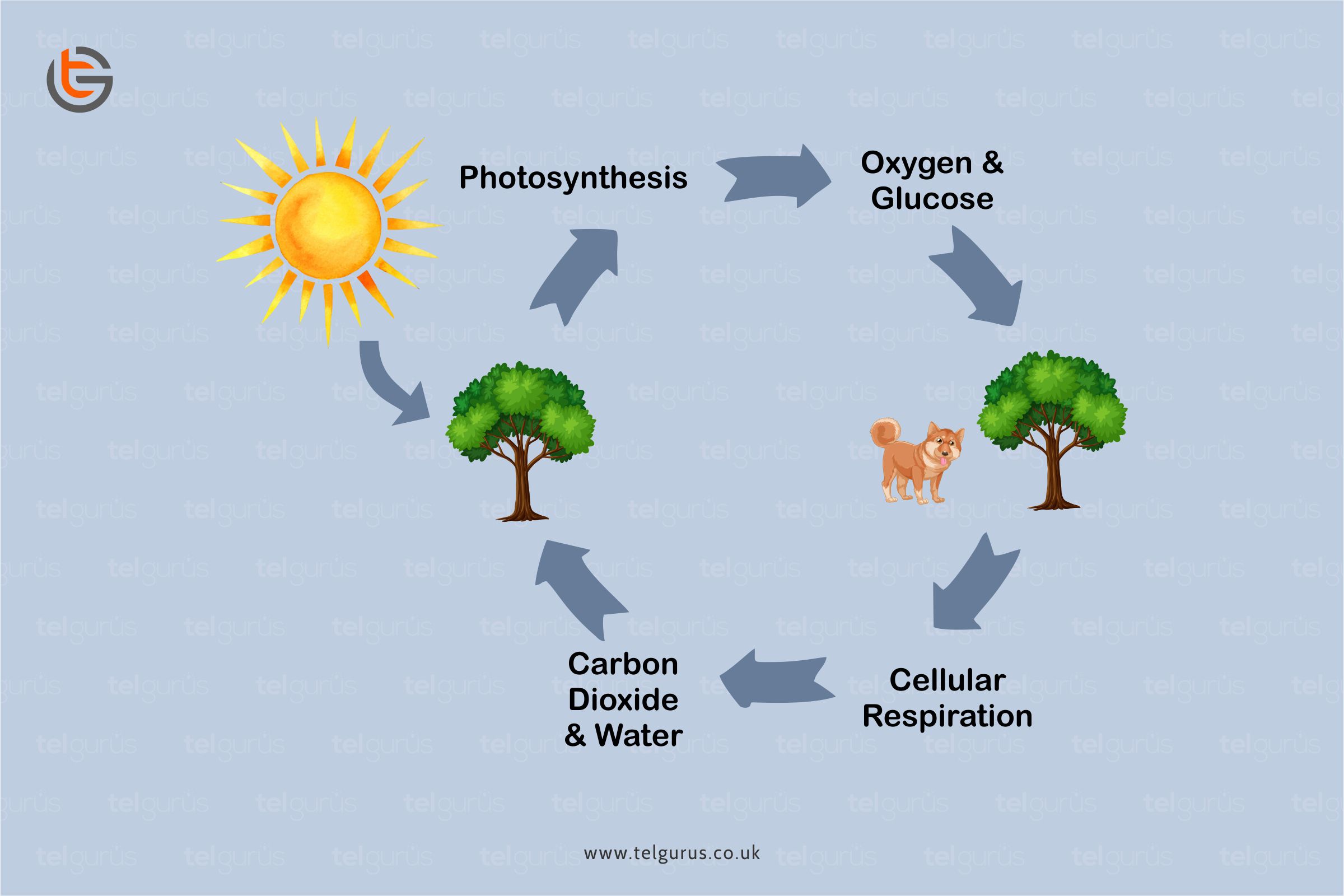
Plants, humans and animals, all depend on the cycle of photosynthesis and respiration for survival.

The oxygen produced by the plants during the process of photosynthesis is what animals and humans inhale for the blood to transport to the cells for respiration.

And the CO2 produced during the respiration process is released from humans or animals and absorbed by the plants to facilitate endow with the energy they require for the development. This is the never-ending cycle that generally sustains life on earth.

Photosynthesis and cellular respiration are the products of a system that are the reactants of the other.


What is photosynthesis?

Photosynthesis is the process that involves the energy used from water, sunlight, and carbon dioxide to produce oxygen and glucose. Plants use the photosynthesis process to produce energy.


What is Respiration?

Respiration uses oxygen and glucose to produce water and carbon dioxide. The process of respiration breaks down the energy for use.

Similarities between Photosynthesis and Respiration

  • Both the processes involve energy production
  • Both the processes involve gases exchange
  • Both the processes take place in the cell organelle that was considered an endosymbiotic organism.
  • Both involve reduction-oxidation reaction and electron transport chain
  • Both the processes synthesize and utilize ATP
  • Both the processes phosphorylation and electron carriers


Similarities explanation

  • Both involve chemical energy production (ATP)

In Photosynthesis, ATP is generated through light energy and utilized to make the organic molecules, whereas, in respiration, ATP is produced by breaking down the organic molecules.

  • Both include ATP production involving an electron transport chain and chemiosmosis.

In photosynthesis, the electrons are donated by the chlorophyll, and protons accumulate within the thylakoid’s lumen, whereas in respiration, electrons are donated by hydrogen carriers. Protons accumulate in the intermembrane space.

  • Both take place in cell organelle.

Photosynthesis occurs in Chloroplast, whereas respiration occurs in mitochondria.


Differences between Photosynthesis and Respiration

Parameters Photosynthesis Respiration
Takes place in It takes place in Algae, plants, and photosynthetic bacteria It takes place in all living organisms.
Function It captures, converts, and stores energy It releases energy
Purpose It converts light energy from the sun that is converted into chemical energy and stored in the glucose bonds Here the chemical energy is stored into the glucose that is released to produce the ATP for cell
Metabolic Process The metabolic process here is anabolic.

Here the energy from ATP NADPH, and CO2 are utilized to produce the glucose molecules.

The metabolic process is catabolic.

Here the glucose is broken down to produce the CO2 and energy in the form of NADH, ATP, and FADH2

Reactants Wate, Light energy and Carbon Dioxide Oxygen and Glucose
Energy source Sunlight Glucose
Location The chloroplast of the plant’s cell Glycolysis takes place in the cytoplasm while the mitochondria are the site of Krebs cycle, ETC
Electron Carriers NADPH NADPH and FADH2
Products Glucose and Oxygen Water and Carbon Dioxide
Equation 6CO2 + 6H2O –––> C6H12O6 + 6O2 C6H12O6 + 6O2 –––> 6CO2 + 6H2O


Bottom Line!

Both respiration and photosynthesis include the energy conversions from one form to another through the biochemical reaction’s series.

Both the processes utilize and produce ATP in reactions, which are carried out on the membranes and controlled by the enzymes. Both the processes involve reaction cycles, including the redox reactions.

Read More – Biology Questions

View More – Useful links for Your Child’s Development 

What is the role of the Nucleus?

The Nucleus is an essential organelle of a cell that acts as a blueprint for how the cells present in the body function. It is responsible for regulating all forms of cellular activities.

Let us get acquainted with the essential information concerning the Nucleus, starting with its definition.

What is Nucleus?

The Nucleus is the most integral component of a cell. It is derived from a Latin word that means “Kernel of a nut.”

It is defined as a double membrane eukaryotic cell organelle, which contains genetic material.


Structure of a Nucleus

The Nucleus is the most evident organelle in a cell that is entirely bound by the membranes. It is the largest organelle that is present in the human body occupying almost 25 percent of the cell volume.

The structure of a nucleus is divided into four main parts. This includes:

  1. The Nuclear envelope

The Nucleus is bound by a double membrane layer which forms the envelope or the capsule.  This envelope’s two layers stay separated from each other by a space often known as the perinuclear space. It generally separates the nucleus’ inner contents from the rest of the cell.

The nuclear envelope’s outer layer is rough due to the ribosome’s presence on its surface. Also, the nuclear membrane has tiny gaps known as the pores that allow the substances’ selective package between the cytoplasm and Nucleus.

Key points of Nuclear envelope

  • It is made up of a membrane phospholipid bilayer having outer and inner membranes.
  • It helps in maintaining the shape of the Nucleus
  • It encloses the nucleoplasm
  • It is connected to the Endoplasmic Reticulum


  1. The Chromatin

The DNA is prearranged in the Nucleus to form chromatin. The chromatin contains the proteins and condenses further to generate the chromosomes. A human cell has a total of 23 pairs of chromosomes.

Key Points of Chromatin

  • The chromatin contains proteins
  • The chromatin condenses to form the chromosomes.


  1. The nucleolus

It’s a well-defined spherical structure that is present within the Nucleus. It is basically the site for the ribosomes’ synthesis and assembly. Here the ribosomes act as protein synthesis sites within the cell.

Key Points of Nucleolus

  • It appears as a darker part of the Nucleus when analyzed with a microscope
  • The Ribosome synthesis takes place here


  1. The nucleoplasm

The nucleoplasm is also known as karyoplasm or nuclear sap. It is a granular, semi-solid substance that comprises several proteins.

The protein fibers generate a crisscross matrix within the Nucleus that helps maintain the structure and the shape of the Nucleus. It is basically the main site for the enzyme activity within the Nucleus.

However, the nucleoplasm appearance may vary during the various phases of a cell cycle.

Along with the proteins, the nucleoplasm also comprises other substances, including RNA, DNA and nucleus.

Key Points of Nucleoplasm

  • Chromatin is the DNA wrapped around proteins
  • It’s a gel-like material that chromatin is stored in


Functions of the Nucleus

The Nucleus performs several essential functions in a cell. The three primary functions of the Nucleus include

  • It comprises the cell’s genetic information in the form of DNA or chromosomes and, therefore, controls cell multiplication and growth.
  • It is also regarded as the site of DNA replication where the formation of an identical copy of DNA occurs.
  • It is responsible for regulating metabolism by synthesizing several enzymes.
  • It is the site for RNA synthesis that acts as a template for the synthesis of several proteins present in the cell.


Bottom Line!

A nucleus is a double membrane organelle consisting of the genetic material and other instructions needed for the cellular process. The Nucleus has two primary functions, including

  • A nucleus is responsible for the hereditary material of a cell.
  • It is also responsible for coordinating several essential activities like cell division, protein synthesis, cell growth, and a host of other vital functions.


Read More – Biology Questions

View More – Useful links for Your Child’s Development 

What is the difference between the pulmonary and systemic circuits of the cardiovascular system?

Our circulatory system comprises the heart, veins, and arteries that convey blood throughout the body. In order to sustain life, the blood must always circulate. It generally carries oxygen from the air. The heart-pumping drives this blood flow through the capillaries, veins, and arteries.

One blood vessel circulates the blood through the lungs for the purpose of gas exchange, whereas the other vessels fuel the rest of the body.


There are two Circulation Types

  • Pulmonary Circulation
  • Systemic Circulation


What is Pulmonary Circulation?

Pulmonary Circulation circulates the blood between the lungs and the heart. It carries the deoxygenated blood to the lungs in order to absorb oxygen and liberate carbon dioxide. This oxygenated blood then flows back to the heart.


What is Systemic Circulation?

Systemic Circulation circulates the blood between the heart and the body. It transforms the oxygenated blood to the cells and then returns the deoxygenated blood to the heart.

Let us understand both the types of circulations and their differences with the help of a few common terms.

  • The heart powers both the circulation types

To begin the systemic Circulation, the heart pumps the blood out of the left ventricle and into the aorta.

Post the blood has supplied the cells throughout the body with essential nutrients and oxygen, it returns the deoxygenated blood to the heart’s right atrium.

And this deoxygenated blood then shoots down from the right atrium to the right ventricle.

Furthermore, the heart then pumps it out of the right ventricle to the pulmonary arteries to initiate pulmonary Circulation. The blood then moves to the lungs, exchanges the CO2 for Oxygen, and returns back to the left atrium.

At last, this oxygenated blood then shoots from the left atrium again to the left ventricle and begins the systemic Circulation.

  • The Circulatory system functions in Tandem with the respiratory system

The respiratory and circulatory systems work together to maintain the body with oxygen and take away carbon dioxide.

Pulmonary Circulation facilitates the external respiration process where the deoxygenated blood flows into the lungs. It absorbs the oxygen from small air sacs and liberates the Carbon dioxide to be exhaled.

On the other hand, Systemic Circulation facilitates internal respiration, where the oxygenated blood flows into the capillaries through the rest of the body.

The blood then diffuses the oxygen into the body cells and absorbs the Carbon Dioxide.

  • The Pulmonary loop transports the blood between the heart and lungs while the systemic loop goes all over the body.

In a pulmonary loop, the deoxygenated blood exits the heart’s right ventricle and passes through the Pulmonary trunk.

The Pulmonary trunk then splits into the left and right pulmonary arteries. These arteries are responsible for transporting the deoxygenated blood to the capillary beds and artioles in the lungs.

There the CO2 is liberated, and oxygen is absorbed. The Oxygenated blood then passes from the capillary beds through the venules in the pulmonary veins.

These pulmonary veins then transport it further to the heart’s left atrium. These pulmonary arteries are the ones that carry the deoxygenated blood, and the pulmonary veins are responsible for taking the oxygenated blood.

Coming to the systemic loop that goes all over the body, the mechanism flows like the oxygenated blood is pumped from the heart’s left ventricle through the aorta, the largest artery present in the body.

The blood transforms from the aorta through the systemic arteries to the arterioles and capillary beds that are responsible for supplying the body tissues.

Here the essential nutrients and oxygen is liberated, and the carbon dioxide and several other waste substances are absorbed.

The deoxygenated blood then travels from the capillary beds through the venules into the systemic veins that feed into the superior and inferior venae cavae, the largest veins present in the body.

And at last, the venae cavae flow the deoxygenated blood to the heart’s right atrium.


Bottom Line!

Both the pulmonary circuits and the systemic circuits of the cardiovascular system perform separate functions and adhere to several differences. However, the essential difference includes:

The pulmonary circuit transports the blood between the lungs and heart. In contrast, the systemic circuit carries oxygenated blood from the heart to the body and then takes the deoxygenated blood from the body to the heart.

Read More – Biology Questions

View More – Useful links for Your Child’s Development 

What are specialised cells?

A human body is made up of several microscopic cells that work in harmony to form the functioning of a human body. However, some of these cells are complex while others are specialized ones.


What are specialized cells?

Specialised cells are the ones that have developed specific characteristics to execute a particular function.

Specialised cells have a particular role to perform and each cell has a different job. And these specialized cells have special features that permit them to perform these jobs.

For instance, the muscle cells that are held together in the bundles pull altogether to make the muscles contraction.

Let us get acquainted with some examples of specialized cells.


Red Blood cells

RBCs, an acronym for Red Blood Cells carry the oxygen around the body. Its specializations include:

They are appropriate for this function because

  • RBCs contain hemoglobin that carries the oxygen molecules
  • RBCs do not have a nucleus that allows them more space to carry oxygen.
  • RBCs are flat disc-shaped giving them a larger surface area and the best chance to absorb as much oxygen as possible.


Nerve Cells

Nerve cells are also specialised cells that transmit electrical signals. Nerve cells are appropriate for this function because

  • Nerve cells are thin and can be almost 1 meter long which means that they can carry the messages up and down the body over large distances
  • Nerve cells have a fatty sheath that surrounds them and this fatty sheath helps the cells in increasing the speed at which the messages can travel.
  • Nerve cells have some branched connections at each end that connect to the other nerve cells and allow them to pass the messages around the body.


Muscle Cells

Muscle cells bring the body’s parts closer. These are appropriate for this function because

  • Muscle cells are generally held together in the form of bundles that pull together to make the muscles contract.

However, being a broad category the muscle cells include different types of muscle cells that each of them perfectly adapt to its function.

  • Cardiac muscle cells: Cardiac muscle cells are specifically for the heart that is branched and join together to generate a net. These muscle cells contract rhythmically and never get tired.
  • Skeletal muscle cells: Skeletal muscle cells are linked to the bones that contract to make the joints bend and bones move.
  • Smooth muscle cells: Smooth muscle cells generate thin muscle sheets like stomach linings. These can also be arranged in the form of rings or bundles like that in the anus.


Sperm Cells

Sperm cells are the necessary cells for human reproduction. These cells are predominantly made up of a nucleus.

These cells are highly mobile since they are required to move to locate an egg for the fertilization process to occur.

The mitochondria present within the sperm cell endows with the energy that is required by the cells to move at much higher rates of speed.


Leukocyte

Leukocyte cells function to keep the body infection-free. These cells are highly flexible and capable of shifting space as required.

Leukocyte cells locate and destroy the microbes within the human body while responding to and treating the infection.

Since these cells must move to the infection site, these are highly capable of pushing through the capillary walls when required to reach the infection’s site.


Bottom Line!

The specialized cells are the ones that are specifically designed to perform the specific functions for which they are intended. We hope that this answer gave you a clear idea about the specialized cells.

Read More – Biology Questions

View More – Useful links for Your Child’s Development 

What is the difference between a receptor and an effector in the nervous system?

The receptors are sensitive to the changes occurring in internal or external environments, whereas the effectors are the glands or muscles that produce action in response to the stimulus.

Both the terms have several similarities and key differences. Let us get acquainted with each one of them.


What is a receptor?

A receptor identifies, detects the stimuli, and converts them into an Impulse.

The receptor generally receives the electric signals. An example of a receptor includes the light receptors in the eye.

Receptors are the cells or a group of specialized cells that can detect some change in the stimulus or environment and generate electrical pulses in the response.

The sense organs are composed of the receptor group that responds to a particular stimulus.

Sensory nerves carry the impulse produced from the stimulus to the central nervous system to process; after processing and interpreting the signals, the central nervous system sends the information to the effectors to generate a response.


What is an effector?

Effectors are the organs that manifest the central nervous systems’ instructions according to the external stimulus. The example of an effector includes a muscle.

Effectors are the body parts like glands and muscles that generate a response to a detected stimulus. These are present in any of the body parts, and motor neurons carry the impulses to effectors.

Once the effectors obtain the impulses, they respond immediately to convert them into actions.

For instance, a muscle contracting to move an arm or a gland liberating a hormone into the blood.


Similarities between receptor and effector

  • Both the effectors and receptors respond to stimuli
  • They convert or generate nerve impulses
  • Information flows from receptors to effectors
  • They work with the central nervous system
  • They are connected to neurons


Understanding the differences between a receptor and an effector in the nervous system

Parameter Receptor Effector
Main action Receptors respond to the stimulus These are the organs that produce a response
Function It sends information to the central nervous system It obeys the commands of the central nervous system
Where it is present It is present in the sense organs only These are present all over the body
Examples include The light receptors in the eye

OR

Ears sound receptors

Muscles contract to move an arm

OR

A gland releases a hormone into the blood.

Definition The receptor detects the stimuli and covers them into an impulse Effector converts an impulse into action.
Detailed Definition It is a cell or a group of cells that are present in the sense organs that are sensitive to a specific type of stimuli like sound, smell, light, heat, taste, or temperature. It is a body part that responds to the stimuli as per the instructions sent from the central nervous system.
Connected to? These are connected to the sensory nerves These are connected to motor nerves.
Present in Sensory organs All over the body
Type of structure A group of cells of a sensory organ A gland or muscle


Bottom Line!

The major difference between the receptor and effector of a nervous system is that a receptor detects the stimuli and converts them to an impulse. In contrast, an effector converts an impulse into action.


Read More – Biology Questions

View More – Useful links for Your Child’s Development 

What is the difference between allopatric and sympatric speciation?

During the evolution process, the formation of distinct and new species is known as speciation.

New species generally form by two primary mechanisms, namely allopatric speciation and sympatric speciation.

Allopatric and sympatric speciation are two mechanisms involved in the new species formation from a pre-existing species. This process of forming a new species from the existing one is known as anagenesis.


What is allopatric speciation?

The allopatric speciation is also called geographical speciation. The difference in the environmental factors leads to the modification in allopatric speciation.

It occurs when two species live in separate environments, and therefore there is no gene flow between the populations.

This will then lead the populations to differentiate as they become adapted to various niches and the environment they are living in.


Understanding it in detail

A specific population can be geographically separated because of some extrinsic barriers like land topography that occurred due to earthquakes, swamps, ice fields, mountains, and deserts.

Once the population is geographically divided, the gene flow ceases between both.

And then, each population becomes genetically diverse due to the different selective pressures of the two different environments they are living in.

Post-separation, the small populations are composed of various allele frequencies as they undergo a founder effect. Therefore, genetic drift and natural selection act differently on the two populations.

Finally, the two diverse genetic backgrounds emerge, leading to new species incapable of interbreeding.


What is sympatric speciation?

In the process of sympatric speciation, the new species evolution occurs from the single ancestral species.

It occurs when two species are living in the same environment. Sympatric speciation is more complicated to understand as the species have occupied the same niche.

However, it may come down to the barriers like post-zygotic or pre-zygotic that prevent the gene flow—for instance, mating at different times of the day.


Understanding in detail

It mainly occurs through polyploidy. Whenever an offspring inherits more than the normal number of chromosomes in the population, this offspring is generally incapable of reproducing with the individuals who include the normal number of chromosomes of the population.

This leads to reproductive isolation within the same population. This process mostly occurs in plants and rarely in animals.

As the plants can self-reproduce, the polyploidy offspring can develop distinct and new generations by themselves.


Understanding the differences between allopatric and Sympatric Speciation

Parameters Allopatric Speciation Sympatric Speciation
Definition The population’s physical isolation due to the extrinsic barrier is known as allopatric speciation. The new species’ evolution from one ancestral species living in a similar habitat is known as sympatric speciation.
Takes place through geographic isolation Yes No
Differentiation mechanism It has natural selection It changes in feeding pattern or its mechanism is polyploidy.
Emerging new species speed It has a slowly emerging new species speed It has a fast-emerging new species speed
Example Darwin’s Finches, Squirrels in the Grand Canyon are a few examples Due to the polyploidy in the plants like tobacco, wheat, maggot flies born on hawthorns and apples, etc.
Common It is common in nature It is common in plants.


Bottom Line!

The major difference between allopatric and sympatric speciation is that the allopatric speciation takes place when the biological population is isolated by some extrinsic barrier leading to an individual’s genetic reproductive isolation.

On the contrary, sympatric speciation takes place when distinct and new species are evolved because of polyploidy.


Read More – Biology Questions

View More – Useful links for Your Child’s Development 

Tel Guru
Tel Guru

Register For The Demo Class

[footer-form]